3D Conformal Radiation Therapy
What is 3D conformal radiation therapy? [lwptoc] 3D conformal radiation therapy, CRT, is a form of cancer treatment that profiles the radiation beams to conform to the shape of the tumour. Previously, radiation beams only correspond to the height and width of the tumour, thereby exposing healthy tissue to radiation. Advancement made in imaging technology,… Read More
Top Doctors For 3D Conformal Radiation Therapy Treatments


3D Conformal Radiation Therapy
What is 3D conformal radiation therapy?
[lwptoc]
3D conformal radiation therapy, CRT, is a form of cancer treatment that profiles the radiation beams to conform to the shape of the tumour.
Previously, radiation beams only correspond to the height and width of the tumour, thereby exposing healthy tissue to radiation. Advancement made in imaging technology, such as 3D CRT, have made it possible to precisely locate and treat the tumour.
3D conformal radiation therapy uses the targeting information to focus accurately on the tumour, while avoiding surrounding healthy tissue. During a 3D conformal radiation therapy, the beams deliver a set dose of treatment to the tumour, which is spread round the surrounding healthy tissue to minimize the entrance and exit of doses to just any one area.
This precise targeting enables the use of higher levels of radiation in treatment. The more the radiation, the more effective the shrinking and killing of tumours. It can also be used to treat noncancerous tumours throughout the body.
What 3D conformal radiation therapy means for you?
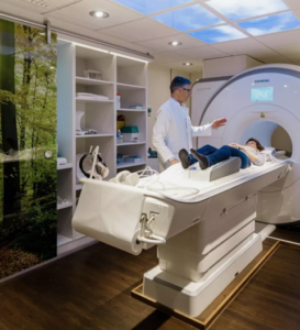
When a 3D conformal radiation therapy is required, the first step is to identify the treatment area using a planning CT scan, magnetic resonance imaging (MRI) scan or positron emission tomography (PET) scan. A personalised treatment plan is then developed using a highly sophisticated radiation therapy software. Each treatment plan will use a number of radiation beams (shaped to the size and targeted at the location, of the particular tumour) from different angles. This means a high dose of radiation is able to be created around the tumour, while sparing a maximum amount of healthy tissue. The individual plan designed is then delivered every day on the linear accelerator (the treatment machine) by radiation therapists.
How does a 3D conformal radiation therapy work?
2D images are gathered from the patient’s CT scans, MRIs or PET scans and fed into a rotating dome-shaped projector plugged into a computer. This dome projects the images as 3D holograms. Holograms are light images which indicate certain characteristics; such as, the size, shape and location of the tumour, and the organs surrounding it.
A 3D conformal radiation therapy uses two devices to create beams:
- Multileaf Collimator: This device is attached to the head of the radiation machine. It contains rows of platelets, separated from each other, known as leaves. The leaves are manipulated to make the initial shape and size of the portal the beams are emitted through.
- Custom-Fabricated, Field-Shaping Blocks: These are made of materials, such as lead, that halts radiation. They are positioned around the portal to further conform the radiation beams to the shape of the tumour.
3D conformal radiation therapy procedure
A 3D conformal radiation therapy session takes near 30 minutes.
The therapist first sets up the treatment plan according to the 3D images of the tumour that has been generated and the info from the simulation.
- The patient lies in the mold on the X-ray table.
- During treatment, beams from a number of directions are accurately matched with the tumour’s height, width and depth.
- The linear accelerator then moves in a circular fashion round the tumour area.
Benefits of 3D conformal radiation therapy
- Improved outcomes from cancer treatment.
- 3D conformal radiation therapy improves control of radiation therapy.
3D conformal radiation therapy side effects
There are a couple of 3D conformal radiation therapy side effects
- Radiation Pneumonitis: An inflammation of the lungs that naturally develops within 2 to 3 months at the start of a radiotherapy. Its symptoms include dry cough, shortness of breath and low-grade fever. Occasionally, it may lead to permanent scarring of the lungs.
- Esophagitis: An inflammation of the oesophagus (the food pipe that runs from the throat to the stomach). This condition generally starts about two weeks after the commencement of treatment and would disappear about 2 to 3 weeks after completion of treatment.
- Mucositis: An inflammation of the oral mucosa – the lining of the mouth, throat and gums. It is associated with dry mouth, thick saliva, sores and difficulty with chewing or swallowing. It’s temporary and goes away within a few weeks of completion of treatment.