Elastography
[lwptoc] Elastography is a medically emerging modality that maps the elastic properties of the soft tissues in the body. In simpler words, elastography is used to detect and determine the stiffness or the softness of the tissue, which accordingly helps in determining the severity of the disease. The hardness or the softness of the tissue… Read More
Top Doctors For Elastography Treatments


Elastography
[lwptoc]
Elastography is a medically emerging modality that maps the elastic properties of the soft tissues in the body. In simpler words, elastography is used to detect and determine the stiffness or the softness of the tissue, which accordingly helps in determining the severity of the disease. The hardness or the softness of the tissue helps provide diagnostic information about the presence or the current status of existing disease. This procedure is known as magnetic resonance elastography, which offers a non-invasive alternative to liver biopsy.
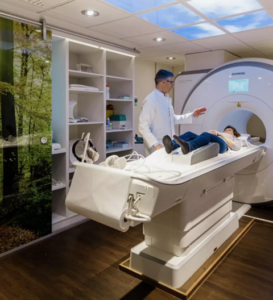
Magnetic resonance elastography (MRE) combines with MRI technology and low-frequency sound waves – with just a regular MRI, doctors can’t figure out whether a patient has fibrosis in the liver. But according to the Mayo Research Clinic, it was found that when used along with the MRI, the MRE technology can determine the stiffness of a liver on a color scale.
How Is An Elastography Done?
Every day, doctors use ultrasound and CT scans to take a look at what is inside the body without having to make an incision. Though these tests are good at showing the size and structure, they don’t show important properties such as tissue stiffness, which is a sign of fibrosis in the liver. However, with the emergence of liver elastography, it is now possible to use sound waves to see if a patient’s liver is harder than it should be and if it is developing fibrosis or not.
During elastography, a drum is placed on the patient’s abdomen. Accordingly, the sound waves move through stiff tissue and supple tissue, each at different rates. A computer analyses the differences and displays the result. The sound waves move the tissues of the upper abdomen and show if a patient has fibrosis in his liver or not – it also shows the location of the fibrosis.
Many liver diseases such as autoimmune hepatitis, hepatitis-C, alcoholism, and other can lead to fibrosis. If the condition is not successfully treated, the disease can progress to cirrhosis, for which the only possible treatment is a liver transplant. MRE is another tool in the fight against the progression of these diseases.
Procedure:
For routine ultrasound elastography, doctors prefer using an intercostal approach to the right lobe of the liver. During this, patients raise their right hand above their heads to increase the intercostal space. A water-based gel (jelly) is applied to the area which is to be studied, helping the transducer securely contact the body.
The radiologist then swipes the transducer across different areas on the skin to find the area of concern.
The doctors then optimize the image to find out the best acoustical window. Then, measurements are taken during breath-hold in a neutral breathing position. These measurements are always taken in the right lobe of the liver as the measurements in the left lobe are often unreliable.
Avoiding the 1st 2 cm from a liver capsule – measurements are taken at an approximate depth of 2cm to optimize the AFRI displacement.
Important guidelines that doctors follow in liver elastography:
1) Avoid Large Vessels And Bile Ducts.
2) ARFI pulse should always be perpendicular to the liver capsule.
3) Avoid Imaging At Vessels.
4) Avoid Imaging depth.
5) Avoid imaging angles.
6) Avoid Imaging < 1.5 cm deep to the liver capsule.
Clinically Proven Benefits Of Liver Elastography
Liver elastography is primarily performed to measure the softness or the stiffness of the tissue and to determine the presence of fibrosis in the liver. In addition to that, elastography also measures the stiffness of the tissue which is influenced by fibrosis. Similarly, it helps in detecting increased hepatic pressure which may be caused by portal hypertension, hepatic congestion, increased blood flow from sugar digestion. Elastography can also map out areas of inflammation in and around the liver. The procedure is minimally invasive and the patient is not exposed to ionizing radiation.
How To Prepare For Ultrasound Elastography?
Though elastography is a non-surgical procedure, there are certain things to know before heading for your examination. During the examination, you will need to remove your clothes and get into a gown for the procedure.
No beverages or sugary drinks should be consumed before the exam as they are known for affecting liver stiffness measurements. Patients are advised to fast for 8 hours before the elastography for better visuals of the gall-bladder and liver tissue. However, these fasting instructions vary from person to person – consult your doctor before going through a liver elastography ultrasound. Claustrophobic patients are allowed to be injected with a mild-sedative before the exam with the doctor’s approval.
Types Of Elastography
- Point Shear Wave Elastography (P-SWE)
In the P-SWE, doctors use the acoustic radial force impulse (ARFI), a low-energy pulse, to generate a shear wave in a small region. The point shear wave elastography is based on real-time imaging allowing doctors to identify and avoid large masses and vessels. Besides, with point shear wave elastography – it is possible to systematically select different parts of a liver for sample.
- 2D Shear Wave Elastography (2D-SWE)
While the 2D-SWE follows the same principle as the P-SWE, it is done multiple times over a larger area. In a 2D-SWE, multiple measurements are taken using the ARFI pulse over a large field of view (FOV). It can either be done as a single image or performed in real-time. Doctors generally prefer real-time imaging to identify and avoid large masses and vessels. 2D-shear wave elastography uses color coding so that the patient can get a general overview of the area. 2D-SWE allows for averaging over a larger area.
Factors Affecting ARFI Measurements
Numerous factors affect the ARFI measurements and can tamper with the elastogram. Here are some of the factors that affect ARFI measurements.
- The amount of tissue displacement is directly dependent on the strength of the ARFI pulse. Therefore, a stronger ARFI pulse may often cause the tissues to displace, hampering your search results.
- The ARFI pulse is attenuated as it traverses tissues. Therefore, measurements taken at a greater depth have less signal to noise (presently 8cm limit on all systems).
- The attenuation is greater with a stiffer liver. Therefore, the measurements can vary accordingly in cases of patients with advanced cirrhosis.
- The ARFI pulse can be refracted by the liver capsule.
- Measurements taken during the patient of scanner motion are inaccurate.
- Measurements taken during inhalation or expiration are inaccurate.
FAQs:
Who interprets the results?
Your transient elastography results, that is the electrogram, will be analyzed by a radiologist, or a doctor trained to supervise and interpret the results of radiology exams.
How do you get the results?
If the liver elastography is conducted at a place other than your doctor’s clinic, then the doctor who carries out the ultrasound elastography will send a report to the doctor who prescribed the elastography. They will then share and explain the results thoroughly to you. At times, the radiologist may contact you directly about your results (if immediate care is needed).
When can you resume your routine activities post-procedure?
As mentioned earlier, transient or ultrasound elastography allows the doctor to examine the internal organs without an incision. It is minimally invasive and therefore, you will be able to resume your activities immediately after the procedure.
Does ultrasound elastography have any side effects?
No harmful effects on humans have been recorded so far in terms of ultrasound elastography.
What should you not carry for the exam?
It is advisable to leave all jewelry and electronic accessories at home before an MRI scan as these items tend to interfere with the magnetic field. These items include things like watches, hair-pins, credit/debit-cards, hearing aid, metal-zippers, pens, pocket knife, eyeglasses, sunglasses, mobile phones, headphones, earplugs, and all accessories with tracking devices.