Ocular Melanoma
Intra-ocular melanoma is a rare type of cancer that forms in the eye. Melanoma is a type of cancer that develops in the melanocytes, the cells responsible for skin, hair, and eye color. Melanoma usually occurs in the skin, but it can develop in the eye on rare occasions, and it is called ocular melanoma. The cancer cells grow in the uvea, which is a layer of tissue located just under the white part of the eye (between the sclera and the retina). Ocular melanoma can also be called uveal melanoma, the uvea has three parts, and each of those parts can be affected by ocular melanoma. Read More
Top Doctors For Ocular Melanoma Treatments


Top Hospitals For Ocular Melanoma Treatments


Ocular Melanoma
Table of contents
Overview
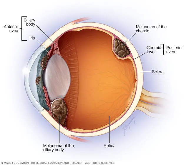
Intra-ocular melanoma is a rare type of cancer that forms in the eye. Melanoma is a type of cancer that develops in the melanocytes, the cells responsible for skin, hair, and eye color. Melanoma usually occurs in the skin, but it can develop in the eye on rare occasions, and it is called ocular melanoma. The cancer cells grow in the uvea, which is a layer of tissue located just under the white part of the eye (between the sclera and the retina). Ocular melanoma can also be called uveal melanoma, the uvea has three parts, and each of those parts can be affected by ocular melanoma. These parts are:
- The iris – This is the colored part of the eye.
- The ciliary body – This is the ring of muscle behind the iris.
- The choroid layer – This is the tissue layer of blood vessels and connective tissue between the sclera and the retina at the back of the uvea.
Ocular melanoma may cause vision issues and could cause serious health issues when it is spread to other organs within the body.
What is the cause of ocular melanoma?
The exact cause of ocular melanoma is unclear. However, it is known that ocular melanoma develops when mutations occur in the DNA of healthy cells in the eye. The mutations cause the cells to grow and multiply in an unregulated manner and not die when they are supposed to. These mutated cells then accumulate in the eye, causing ocular melanoma.
What are the risk factors of ocular melanoma?
The risk factors include:
- Age – The chance of developing ocular melanoma rises as one gets older.
- Light eye color – individuals with light-colored eyes, like blue or green eyes, have a greater risk of ocular melanoma.
- Skin color – white people have a greater risk of developing ocular melanoma than people of other races.
- A family history of ocular melanoma – individuals that have a family member that has suffered from ocular melanoma have a higher risk of developing it.
- Exposure to UV rays – though unclear how, some evidence suggests that exposure to UV rays, like rays from the sun or tanning beds, increases an individual’s risk of developing ocular melanoma.
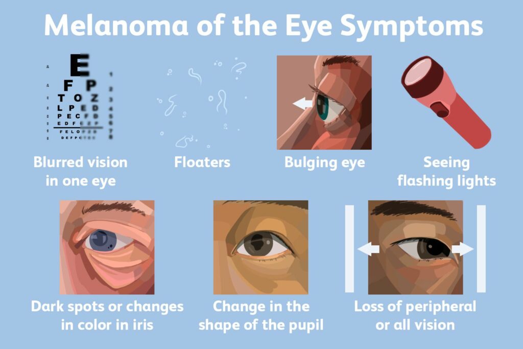
What are the symptoms of ocular melanoma?
:max_bytes(150000):strip_icc()/VWH_Illustration_Melanoma-of-the-Eye-Symptoms_Illustrator_Laura-Porter_Final-d7402eba72c04c02aaed226afae0411c.jpg)
In some cases, ocular melanoma does not present with any symptoms, or the symptoms may be difficult to recognize because the cancer occurs in the part of the eye that is not visible. Some general symptoms that may, however, occur include:
- Blurred vision
- Dark spots on the iris
- Blind spots or reduced field of vision
- Displaced eye lens
- Double vision
- Complete vision loss
- Pain
- Retinal detachment
- Flashes of light in the field of vision
- Alterations in the shape of the pupil
- Changes in the position of the eye in its socket.
- Increasing pressure in the eye.
- Spreading to other organs like the lungs, bones, liver, etc.
How is ocular melanoma diagnosed?
The diagnosis of ocular melanoma often occurs during a routine eye examination. The health care provider will check the medical history and also the presenting symptoms. During an eye exam, the individual’s vision is checked and looked inside the eye. The pupils may be dilated in order to examine the back of the eye. Other additional tests that may be used to confirm a diagnosis include:
- Ultrasound – this is where sound waves are used to visualize the eye in order to measure the size of the eye structures and check for tumors.
- Fluorescein angiography – this is where a special dye is injected into the individual to help examine for any blockages or leakages in the eye.
- Biopsy – in some rare cases where the tests do not give a definite answer, some tissues from the eye may be collected for laboratory examination.
After the diagnosis, more blood and imaging tests may need to be carried out in order for cancer to be staged. Staging is the process of determining the level of spread of cancer.
What is the treatment for ocular melanoma?
If the ocular tumor is not cancerous, it might not need to be treated immediately. The doctor, however, may want to check if it grows or causes any issues regularly; this is called watchful waiting. Ocular melanoma that is caught early can be successfully treated. The treatment plan will also depend on the location of the tumor, the size, and thickness of the tumor, the staging of the tumor, and the age and health condition of the individual. Some of the ways of treatment include:
- Radiation – this is where beams of X-rays are used to destroy tumors. The radiation may be administered by external or internal radiation therapy. External radiation therapy directs the beams from outside the body. Special techniques like proton beam radiation which limits the damage to the eye and brain tissue, may be used. Internal radiation therapy implants the seeds of radiation near the tumor inside the eye. This technique is called radioactive plaque therapy.
- Lasers – Infrared light may be used to kill a small tumor and seal off a blood vessel to prevent the cancer’s spread. This usually involves sending a low-powered laser beam through the pupil to destroy the cancer cells without damaging the rest of the structures in the eye.
- Surgery – this can be done in two ways, they are external resection and enucleation. External resection is done to remove the tumor and some healthy tissues, while enucleation is where the eye and some parts of the optic nerve are removed. Some degree of vision loss is a risk in each type of surgery.
FAQ
Most of the risk factors cannot be altered, like skin color and age. The most significant way to reduce the risk of ocular melanoma is regular eye check-ups by an ophthalmologist. Catching the condition early greatly improves the chances of successful treatment.
It has been proven that ocular melanoma spreads in about 40-50% of cases. Choroid melanoma and ciliary body melanoma are more likely to spread than iris melanoma. Also, in about 90% of the cases that spread, they usually spread to the liver.