Congenital Uterine Anomalies
Congenital uterine anomalies are malformations of the uterus that originate during embryonic life. These uterine anomalies occur only in about 5% of women but have been indicated in up to 25% of women that experience miscarriages or premature baby deliveries. A congenital uterine anomaly occurs when a woman’s uterus develops differently from most women. Normally,… Read More
Top Doctors For Congenital Uterine Anomalies Treatments


Top Hospitals For Congenital Uterine Anomalies Treatments


Congenital Uterine Anomalies
Table of contents
Congenital uterine anomalies are malformations of the uterus that originate during embryonic life. These uterine anomalies occur only in about 5% of women but have been indicated in up to 25% of women that experience miscarriages or premature baby deliveries. A congenital uterine anomaly occurs when a woman’s uterus develops differently from most women.
Normally, when a woman is in the womb, her uterus develops as two separate halves that fuse before she is born. Most women find out that their uterus is different when they go for an ultrasound scan after a miscarriage, bleeding, or infertility problems. The effect these abnormalities have on pregnancies is not clear, but women who have found out about the differences say that it helps them better prepare for pregnancy. These abnormalities may not hinder pregnancy, but they may significantly reduce the ability to carry a pregnancy to full term.
What are the types of congenital uterine abnormalities?
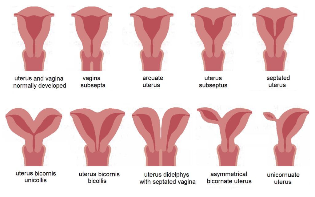
Defects of the uterus may include:
Separate uterus
The uterus in this instance appears entirely normal from the outside, yet an internal septum divides it into two distinct parts. This septum is of varying sizes and thickness and separates the uterus into two endometrial cavities. This is the most diagnosed abnormality making up about 45% of the cases. Women with this condition are likely to have issues with conception. Preterm birth and early miscarriage are also more likely. They may also be advised to have a C-section because the position of the fetus is also affected.
Bicornuate uterus
In this condition, the uterus has an external indentation or groove marking its division internally into different endometrial cavities. The two halves of the uterus will appear almost completely separate except for the lower part. This makes up about 25% of all congenital abnormality cases. Women with this condition have no extra difficulty in conception or early stages of pregnancy but there is a slightly higher risk of miscarriage and preterm birth. It can also affect the baby’s position later in the pregnancy so a cesarean section may be carried out.
Arcuate uterus
The uterus looks normal here but the internal surface of the single endometrial cavity shows a shallow grove of about 1cm or less, it has a slight dip at the top. This condition has no increased risk of early miscarriages or preterm birth, but it may increase the risk of late miscarriage.
Unicornuate uterus
In this condition, only half of the uterus develops from a single Mullerian duct. This condition is rare, it usually indicates that the uterus is half the size of a normal uterus. In this condition, there is an increased risk of an ectopic pregnancy, late miscarriage, or preterm birth. This condition is more common in infertility cases as well.
Uterus didelphys
Here, the two halves of the uterus develop completely separate, with each side having its cavity. It can affect the uterus, the cervix, and sometimes the bladder, vagina, vulva, and urethra. Women with this condition do not have difficulties with conception, with only a slightly increased risk of preterm birth.
Uterine agenesis
This is where the uterus does not develop at all. It occurs in about 10% of all cases of uterine abnormalities
What is the cause of the condition?
Most of these anomalies have unknown reasons. In more than 90% of the cases, the chromosome pattern is normal. Also, there have been no established risk factors or preventative measures.
How is this condition diagnosed?
As stated earlier, most women do not know that they may possess an abnormality until they become pregnant or suffer from infertility. This is because the condition does not have any physical symptoms. They are usually discovered after an ultrasound scan following a miscarriage or in investigating the cause of infertility. It may even be discovered during investigations into gynecological issues like abnormal bleeding. At this point, they may want to consider surrogacy with a surrogate that can carry the pregnancy to term.
While routine scans will usually reveal didelphic or unicornuate uterus, they will not usually reveal the milder forms of uterine abnormality. Further investigations can include the following:
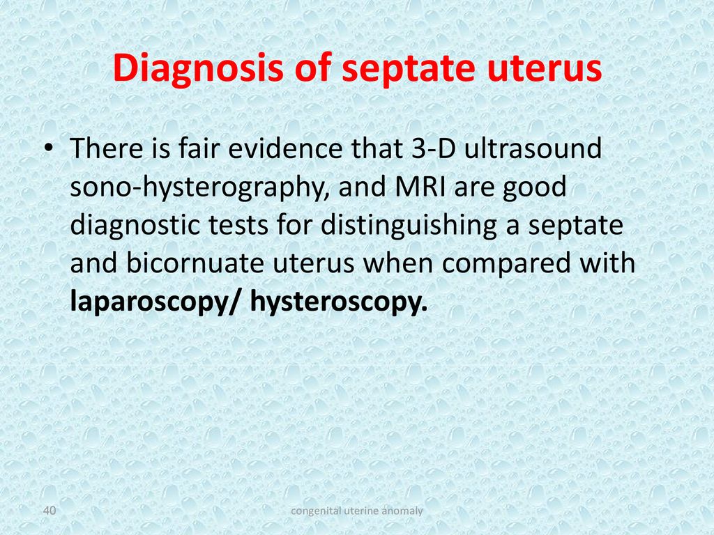
- Hysteroscopy – This procedure involves placing a small camera through the cervix into the uterine cavity. Fluid is then flushed into the uterus to enable the viewing of the whole cavity. This procedure enables the visualization of any abnormality like fibroids and polyps, as well as the shape of the uterus. A few risks are associated with hysteroscopy, they are bleeding, infection, cervical damage, and perforation of the uterus. These risks are rare, however. After the procedure, it is normal to experience cramping and light spotting for a few days.
- Laparoscopy – This is a surgical procedure that allows a surgeon to access the inside of the uterus. A camera is inserted through the abdominal wall to view the uterus, the fallopian tubes, and the ovaries.
- Pelvic ultrasound scan – This is where ultrasound waves are used to visualize the uterus. A transvaginal ultrasound is preferred as the waves are closer to the uterus and the ovaries. This is done by inserting the probe into the vagina in order to view the uterus and the ovaries. Although the procedure may be uncomfortable, it is very brief.
What are the treatment options for Congenital Uterine Anomalies?
This depends on the type of abnormality that is present. While surgical interventions may seem ideal, they can lead to eventual infertility and problems during pregnancy. A gynecologist will usually consult with the patient and discuss the options that they have if the abnormality was discovered before a pregnancy. Overseas medical treatment where hospitals are better equipped will help diagnose these conditions quickly. The aid of a trusted medical travel agency or meditour agency can be sought to find good hospitals for medical tourism.
If the diagnosis is during pregnancy, the patient is placed into the care of an obstetrics team. They will have extra care, scans, and more hospital visits to care for the baby throughout the pregnancy. The healthcare team will also consult with the patient if their baby ends up in the wrong position in later pregnancy. They will recommend a C-section but the option lies on the patient. Septate uteri are usually repaired in order to improve the chances of pregnancy, but other cases are usually left as they are. In cases where obstruction of the female genital tract is present, surgical repair may result in permanent infertility.
In cases where these anomalies are diagnosed before pregnancy, surrogacy can be used. The embryo can be harvested from the mother, and a surrogate can be used to carry the pregnancy to term.